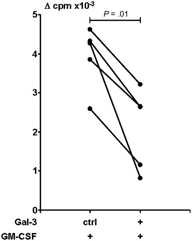
Figure 5.
Effect of galectin-3 (Gal-3) on antigen-presenting cells (APCs) on the ability to present antigen to T cells from leprosy patients. T-cell proliferation using APCs previously treated with granulocyte macrophage colony-stimulating factor in the presence of Gal-3 or control buffer as in Figure 4 are shown. In triplicate, CD1b-restricted T cells and APCs (1:1) were cultured in the presence or absence of mycobacterial antigen (M. tuberculosis sonicate or mycobacterial lipomannan) for 3 days, and proliferation was measured by 3H thymidine incorporation. Proliferative response was reported as average of counts per minute (cpm) of wells in the presence of antigen minus the average of cpm of corresponding wells in medium alone (Δ cpm). T cells cultured with medium alone–derived APCs proliferated at a mean of 65 Δ cpm (range, 2–207 cpm). Shown are results from 5 experiments, 4 donors. Statistical analysis was performed with paired t test.

