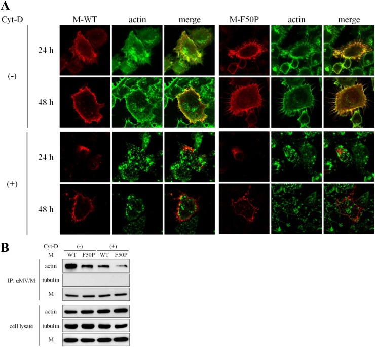
Fig 3.
Tight association of the M-WT protein with F-actin. (A) Colocalization of the M proteins with F-actin. HeLa cells were transfected with a plasmid expressing the M-WT or M-F50P protein. At 12 h posttransfection, culture medium was replaced by medium including Cyt-D or DMSO. At 24 or 48 h posttransfection, the cells were fixed, permeabilized, and incubated with the mouse monoclonal antibody against the M protein followed by Texas Red-conjugated goat anti-mouse IgG. F-actin was visualized with FITC-conjugated phalloidin. Observation was performed under a confocal microscope. (B) Efficient coprecipitation of actin with the M-WT protein. 293T cells were transfected with a plasmid expressing M-WT or M-F50P protein. At 12 h posttransfection, culture medium was replaced by medium including Cyt-D or DMSO. The cells were incubated for a further 12 h and then solubilized with detergent solution after the incubation with cross-linking reagent DSP for 2 h. Proteins immunoprecipitated with the anti-M antibody (IP) as well as whole-cell extracts (cell lysate) were subjected to immunoblot analysis. The M protein, actin, and tubulin were detected using mouse monoclonal antibody against MV M protein, mouse monoclonal antibody against β-actin, and rabbit polyclonal antibody against β-tubulin as the first antibodies, respectively.

