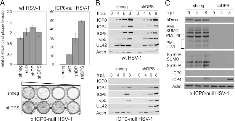
Fig 3.
Consequences of triple depletion on ICP0-null HSV-1 replication in HepaRG cells. (A) Relative efficiency of plaque formation of HSV-1 in depleted and nondepleted cells. Cells were infected with HSV-1 strain in1863 and ICP0-null HSV-1 strain dl1403/CMVlacZ at sequential dilutions. At 24 h postinfection, cells were fixed and stained for β-galactosidase expression. Bars represent mean relative plaque-forming titers in depleted compared to nondepleted (shneg) control cells in three independent experiments. A typical example of a stained plaque dilution series is shown at the bottom, with plaques shown as dark spots. (B) Kinetics of viral protein expression in depleted and nondepleted cells. Cells were infected with wt or ICP0-null HSV-1 at an MOI of 2, and cell lysates were sampled at 0, 4, 6, and 8 h postinfection (h p.i.). Samples were resolved on a 7.5% polyacrylamide gel, and membranes were probed using anti-ICP0 (11060), anti-ICP4 (58S), anti-ICP8 (ab20194), anti-UL42 (Z1F11), and anti-VP5 (DM165) antibodies. The loading control was antiactin serum A5060. (C) The shRNAs in triple-depleted cells are able to overcome any interferon-induced increased expression of PML, Sp100, and hDaxx that might occur during ICP0-null mutant HSV-1 infection. Control (shneg) and triple-depleted (shDPS) cells were infected with ICP0-null mutant HSV-1 at an MOI of 2, and samples were then harvested at the indicated time points and analyzed by Western blotting, as described in the legend of Fig. 2A.

