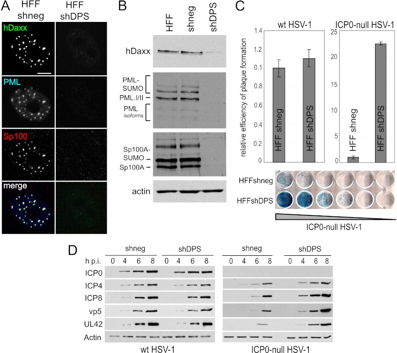
Fig 4.
Triple depletion in HFs improves replication and gene expression of ICP0-null mutant HSV-1. HFs were transduced with a lentivirus expressing shRNA with no cellular target (shneg) or targeting hDaxx, PML, and Sp100 (shDPS). (A) Verification of depletion by immunofluorescence. Cells were simultaneously stained with rabbit anti-hDaxx 07-471 antibody, mouse anti-PML MAb 5E10, and rat anti-Sp100 r26 antibody. Secondary antibodies are described in the legend of Fig. 2. Bar, 10 μm. (B) Immunoblot analysis of extracts from the generated cell lines demonstrating depletion of the respective proteins. Antibodies used for detection are described in the legend of Fig. 2. (C) Relative efficiency of plaque formation in depleted and nondepleted HFs. Cells were infected with HSV-1 strain in1863 and ICP0-null HSV-1 strain dl1403/CMV/lacZ at sequential dilutions. At 24 h postinfection, cells were fixed and stained for β-galactosidase expression. Bars represent mean relative efficiencies of plaque formation determined by calculating virus titers in depleted compared to nondepleted (shneg) cells in three independent experiments. A typical example of a stained plaque dilution series is shown at the bottom, with plaques stained in blue. (D) Kinetics of viral protein expression in depleted and nondepleted cells. Cells were infected with wt or ICP0-null HSV-1 at an MOI of 2, and cell lysates were sampled at 0, 4, 6, and 8 h postinfection (h p.i.). The samples were analyzed as described in the legend of Fig. 3.

