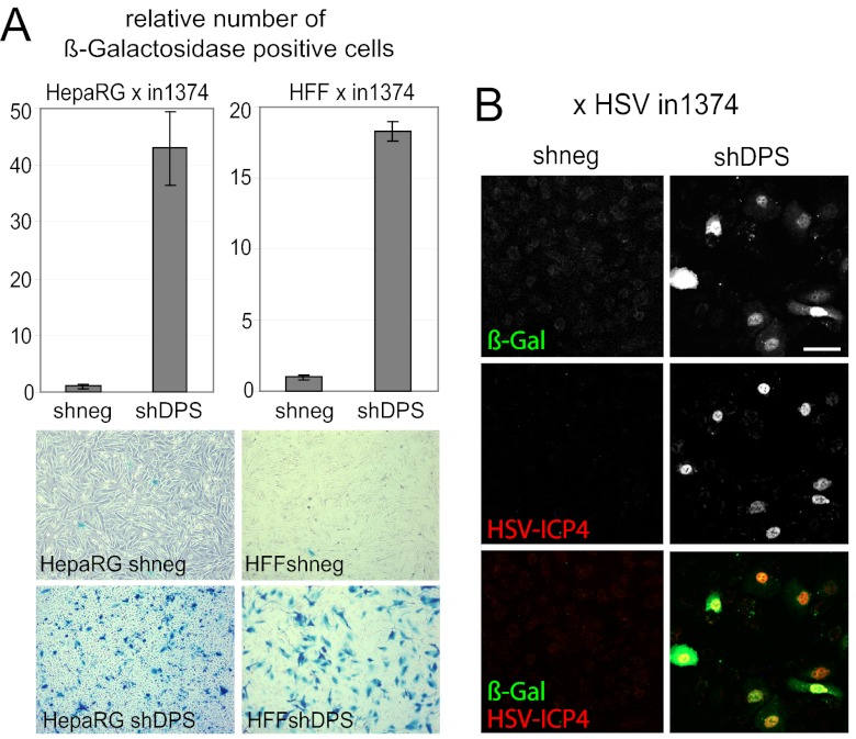
Fig 7.
Establishment of quiescent infection in triple-depleted HFs and HepaRG cells. (A) Cells were infected with HSV-1 strain in1374 and incubated at the restrictive temperature of 38.5°C, and at 24 h postinfection, they were then stained for β-galactosidase activity. (Top) Relative cell numbers of β-galactosidase-expressing cells in HepaRG cells and HFs transduced with a lentivirus expressing shRNAs with no cellular target (shneg) or targeting hDaxx, PML, and Sp100 (shDPS). Bars represent mean values obtained from three independent experiments. (Bottom) Images of the respective stained cell monolayers at 24 h postinfection. (B) Triple-depleted HepaRG cells were infected with in1374 as described above for panel A and then fixed at 24 h postinfection and stained for immunofluorescence analysis with anti-β-galactosidase and anti-ICP4 (r74) antibodies. Bar, 100 μm.

