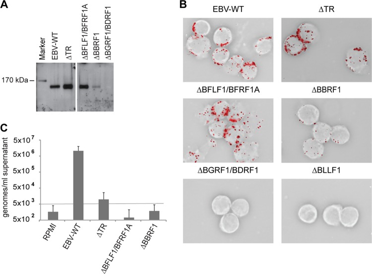
Fig 5.
Purification and analysis of DNA-free defective virus particles. (A) Virus particles from 1 ml of the indicated supernatants were pulled down using gp350-specific antibodies and protein extract thereof were separated through an acrylamide gel. Blotted proteins were incubated with a rabbit antiserum specific for BNRF1 tegument protein. Wild-type viruses were used as a positive control. (B) Elijah B cells were incubated with 1 ml of the various virus supernatants, and bound particles were visualized by immunostaining using a gp350-specific antibody. Wild-type virus was used as a positive control and gp350 knockout mutant virus (ΔBLLF1) as a negative control. (C) The viral DNA content per ml of purified supernatant was determined by qPCR analysis using an EBV-specific probe. This assay used RPMI and wild-type viruses as negative and positive controls, respectively. The lower limit of quantitative measurement is 5 × 10³ DNA molecules per ml of supernatant. The mean results and standard deviations from eight independent experiments are given.

