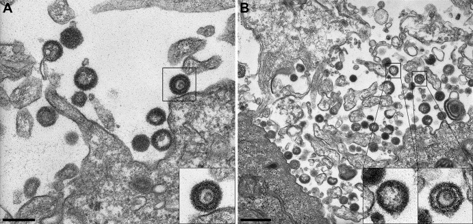
Fig 7.
Electron microscopy of cells infected with a PrV UL32 deletion mutant. (A) VLPs and LPs are released at the cell membrane of infected cells. The inset represents an example of a VLP (bar, 250 nm). (B) Multiple LPs and VLPs are present in the extracellular space. The inset on the left shows an LP and that on the right a VLP (bar, 500 nm).

