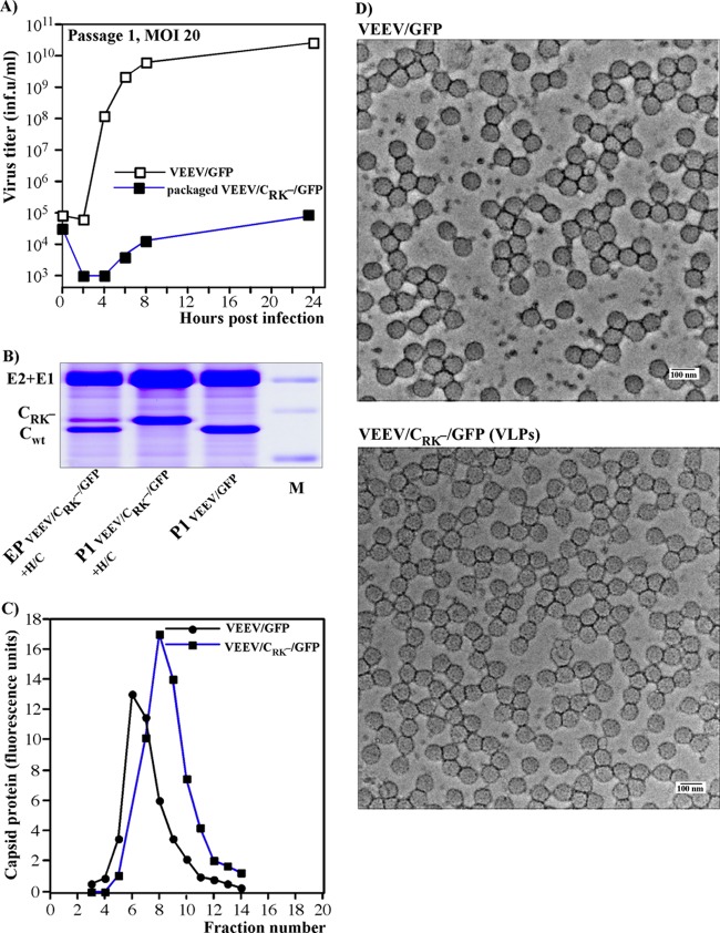
Fig 3.
Cells containing replicating VEE PIV genomes produce noninfectious VLPs. (A) Subconfluent BHK-21 cells in 6-well Costar plates were infected with viral or PIV genome-containing viral particles, generated by coelectroporation of VEEV/CRK−/GFP and H/C helper genomes into the cells, at an MOI of 20 infectious units/cell. Media were replaced at the indicated times postinfection, and infectious titers were determined as described in Materials and Methods. (B) Analysis of viral particles released from cells coelectroporated with in vitro-synthesized VEEV/CRK−/GFP PIV and H/C helper RNAs or infected at an MOI of 20 infectious units/cell with particles containing the packaged PIV genome or with VEEV/GFP. Media were harvested at 24 h postelectroporation or -infection, and particles were pelleted by ultracentrifugation and analyzed by SDS-10% PAGE followed by Coomassie blue staining. Each line on the gel contains an aliquot corresponding to 3 ml of harvested medium. (C) Analysis of the density of particles released by cells either infected by virus or infected by PIV genome-containing particles. Analysis was performed by ultracentrifugation of sucrose density gradients as described in Materials and Methods. (D) The genome-containing wt viral particles and VLPs formed by the replicating VEEV/CRK−/GFP PIV genome were concentrated from serum-free medium by use of centrifugal Ultracel-100K filters (see Materials and Methods for details). These samples were used directly for negative staining and EM analysis (see Materials and Methods for details).

