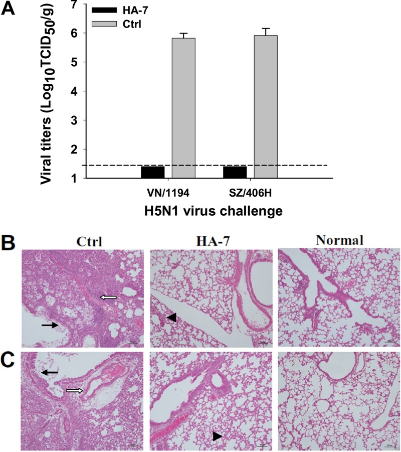
Fig 3.
Virus titers and histopathological changes in the lungs of MAb-treated mice challenged with H5N1 viruses. HA-7-injected mice were challenged with 5 LD50 of clade 1 strain VN/1194 and clade 2.3.4 strain SZ/406H of H5N1, and lung tissues were collected at day 5 postchallenge. (A) Detection of viral titers of VN/1194 and SZ/406H H5N1 viruses in collected lung tissues. Mice injected with SARS-CoV RBD-specific MAb were used as the negative control (Ctrl). The data are expressed as means ± standard deviations of virus titers (log10 TCID50/g) of lung tissues from six mice per group. The limit of detection is 1.5 log10 TCID50/g of tissues, as shown by the dotted line. The figure shows representative data from two independent repeat experiments. (B and C) Evaluation of histopathological changes in collected lung tissues. Lung tissues from the mice injected with a MAb to the RBD of SARS-CoV and those from uninfected mice were used as the negative control (Ctrl) and normal control (Normal), respectively. Lung tissues were stained with H&E and observed by light microscopy (magnification, ×100). Shown are representative images of histopathological changes from two independent experiments with six mice per group challenged with VN/1194 (B) and SZ/406H (C) H5N1 viruses, respectively. Serious histopathological damage was seen in the control mice, with bronchial epithelial cell degeneration, necrosis, and desquamation (solid arrows). Large amounts of exudates and severe edema with infiltration of lymphocytes and mononuclear cells were also seen around blood vessels (open arrows). However, the HA-7-immunized mice showed only mild interstitial pneumonia changes with focal broadening interstitial spaces and lymphocytic infiltration (arrowheads).

