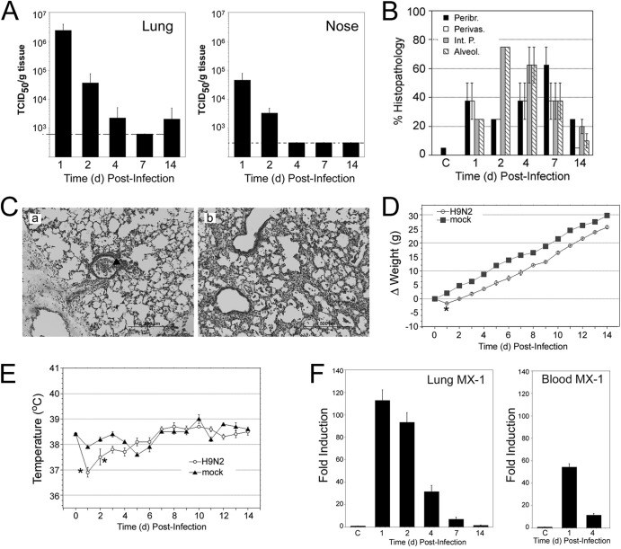
Fig 3.
Characterization of infection of cotton rat with H9N2 A/guinea fowl/Hong Kong/WF10/1999. (A) Lung and nose viral replication after infection with 107 EID50/rat. (B) Quantification of lung pathology for 4 relevant parameters of damage: peribronchiolitis, perivasculitis, interstitial pneumonia, and alveolitis. Pathology peaked at 2 to 7 dpi and was still detectable at 14 dpi. (C) Lung pathology was characterized by strong accumulation of cell debris and granulocytes in the airways, forming plugs at 1 dpi (panel a, arrow). The major airway infiltration is resolved by day 4 (panel b), and the pathology progresses to a strong interstitial pneumonia accompanied by alveolitis. Magnification, is ×100. (D and E) Weight (D) and temperature (E) changes were recorded at different days postinfection. *, P < 0.05. (F) mRNA expression of the type I interferon-inducible gene MX-1 in lung and blood measured at the indicated dpi. C, group of mock (allantoid)-inoculated animals.

