
Fig. 7. Displacement of streptavidin from a 5′-biotinylated oligonucleotide by Hel-1a, R393A, and F438A.
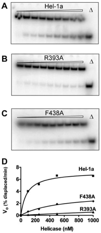
HCV helicase was incubated with a [32P]oligonucleotide with biotin at its 5′-end bound to streptavidin. When ATP is added to the reaction, streptavidin is displaced from the oligonucleotide leading to a shift in the electrophoretic mobility of the [32P]DNA. Autoradiographs of non-denaturing gels are shown of reactions containing 100 nm of Hel-1a (A) R393A (B) or F438A (C). Reactions were analyzed before ATP addition (lane 1) and after 1, 3, 6, 9, 12, 15, 20, and 30 min (lanes 2–9). Also shown are the products after boiling (Δ, lane 10). D, initial rates of streptavidin displacement that were measured in reactions containing various amounts of Hel-1a (squares), R393A (triangles), and F438A (diamonds). Data were fit to Equation 3.