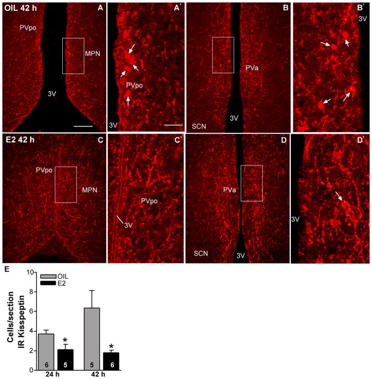
Figure 11.
Immunoreactive kisspeptin in the POA from oil- and E2-treated females. Representative photomicrographs of immunoreactive kisspeptin in coronal sections through the POA from rostrally (A,C) to caudally (B,D) in oil-treated (A,B) and E2-treated (C,D) animals (42 hours after treatment). The boxed areas in A–D are amplified in A′–D′, respectively, to illustrate immunoreactive cells (arrows). E: Quantitative analysis of the effects of E2 on the number of kisspeptin-immunoreactive cells in POA during negative (24 hours) and positive (42 hours) feedback. *P < 0.05; n = 5–6, two-tailed Student’s t-test. Scale bars = 165 μm in A (applies to A–D); 40 μm in A′ (applies to A′–D′). [Color figure can be viewed in the online issue, which is available at wileyonlinelibrary.com.]

