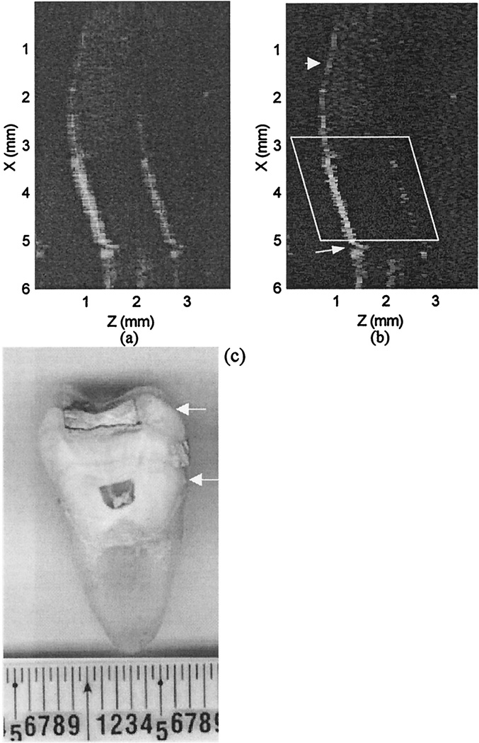
Fig. 7.
(a) Image of a tooth with a metal filling at the surface. (b) CLEANed image. (c) Microscopic picture of the sectioned tooth. The spatial scan range is indicated by the two arrows in (c). The portion of the –air-enamel interface that is delineared well after use of CLEAN is pointed to by the arrow at the top of (b). The indented portion of the air–enamel interface corresponds to a cavity and is pointed to by the the longer arrow at the bottom of (b). The boundary and the internal aspects of the metallic restoration (within the frame) are more clearly seen after CLEAN is applied.

