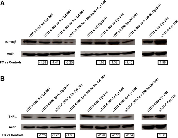
Figure 5.
Expression of IGF1Rβ and TNFα proteins is regulated by miR-296-3p and miR-298-5p in αTC1-6. (A) Western analysis of IGF1Rβ in (1) untreated αTC1-6 transfected for 24 h with (i) scramble molecules (NC); (ii) mimics of miR-296-3p; (iii) mimics of miR-298-5p; (iv) mimics of both miR-296-3p and miR-298-5p (left); (2) αTC1-6 transfected for 24 h with (i) scramble molecules (NC); (ii) mimics of miR-296-3p; (iii) mimics of miR-298-5p; (iv) mimics of both miR-296-3p and miR-298-5p and treated with cytokines for further 24 h (middle); (3) αTC1-6 treated with cytokines for 24 h and their matched untreated controls (right). (B) Western analysis of TNF-α performed in the same experimental conditions as (A). β-Actin signal was used to normalize the data. Numbers below Actin blots represent fold change expression values relative to matched controls.

