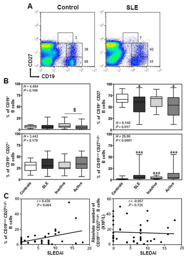
Figure 1.
Increased frequency of circulating plasma cells in SLE patients. (A) Double staining with CD19 and CD27 was performed on PBMC to delineate naive (CD19+CD27-) B cells, memory (CD19+CD27+) B cells and plasma (PC, CD19lowCD27high) cells. Thresholds and fluorescence gates used for the statistical evaluation of CD27-, CD27+ and CD27high B lymphocytes are indicated as well as the corresponding frequency of these subsets among total B cells. Expression of CD27 on CD19+ peripheral B cells is shown for a representative healthy blood donor and a patient with active SLE. (B) Comparison of the frequencies of total B cells among PBMC and of the different B-cell subpopulations from SLE patients (n = 41), distributed according to disease activity, i.e. inactive (n = 17) versus active (n = 24), and from healthy individuals (n = 45). The proportions were determined by flow-cytometric analysis as shown in A. Results are depicted as box plots, with median (horizontal line within each box) and 10th, 25th, 75th, and 90th percentiles (bottom bar, bottom of box, top of box, and top bar, respectively). Kruskal-Wallis H test and associated P values are indicated. *P < .05 and ***P < .0005 compared with control B cells. §P < .05 compared with B cells from patients with inactive SLE (as determined using the Mann–Whitney U-test). (C) Frequency (left panel) or total number (right panel) values obtained within CD27high PC for each SLE patient were plotted as a function of SLEDAI scores. Correlation factor (r, Spearman’s) and P value are indicated.

