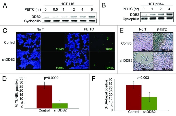
Figure 2. PEITC induces expression of DDB2-independent of p53. (A) HCT116 cells were treated with PEITC for indicated time point. Total RNA was analyzed by semi-quantitative PCR for the level of DDB2. Cyclophlin was used as a loading control. (B) HCT116 p53−/− cells were treated with PEITC for indicated time points. Total RNA was analyzed by semi-quantitative PCR for the level of DDB2. Cyclophlin was used as a loading control. (C) HCT116 cells expressing control shRNA or DDB2 shRNA were treated with PEITC for 12 h. Next day, cells were analyzed for apoptosis by TUNEL assay. Representative images of HCT116 cells expressing control shRNA or DDB2 shRNA stained for TUNEL assay after PEITC treatment. (D) TUNEL positive cells were counted from at least 10 fields of triplicate plates. A quantification of TUNEL positive HCT116 cells after PEITC treatment is shown. (E) HCT116 cells expressing control shRNA or DDB2 shRNA were treated with PEITC for 4 h. After 48 h, cells were analyzed for SA β gal activity. Representative images of HCT116 cell expressing control shRNA or DDB2 shRNA stained for SA β gal after PEITC treatment. (F) SA β gal positive cells were counted from at least 10 fields of triplicate plates. A quantification of SA β gal positive HCT116 cells after PEITC treatment is shown.

An official website of the United States government
Here's how you know
Official websites use .gov
A
.gov website belongs to an official
government organization in the United States.
Secure .gov websites use HTTPS
A lock (
) or https:// means you've safely
connected to the .gov website. Share sensitive
information only on official, secure websites.
