
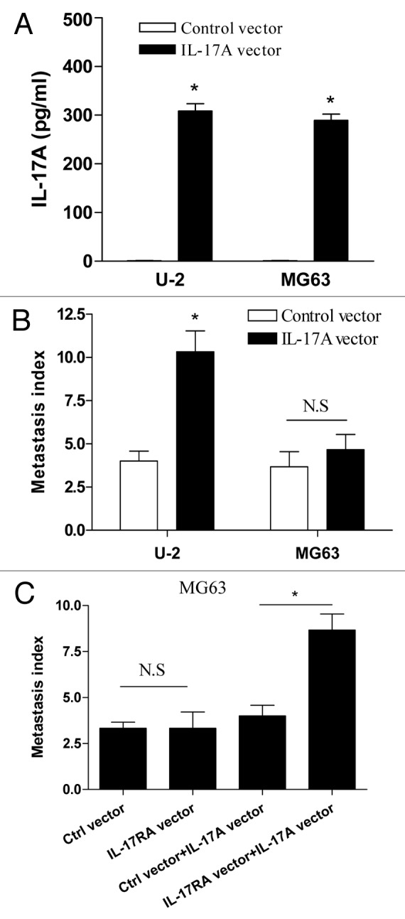
Figure 5. IL-17A/IL-17RA interaction enhanced the metastasis of OS cells in vivo. (A) 1 × 10⁶ U-2 cells or MG63 cells stably transfected with IL-17A expression vector or control vector were incubated at 6-well plates respectively. After 48 h, the concentration of supernatant IL-17A was determined by ELISA assay. (B) Groups of eight nude mice were challenged with 2 × 10⁶ of U-2 cells or MG63 cells which were stably transfected with IL-17A expression vector or control vector, respectively. (C) Groups of eight nude mice were challenged with 2 × 10⁶ G63 cells which were stably co-transfected with IL-17A expression vector and IL-17RA expression vector, or the corresponding control vector, respectively. After 30 d, the metastatic index to lung was determined. Each bar represents the means (± SD) in each group. *p < 0.05