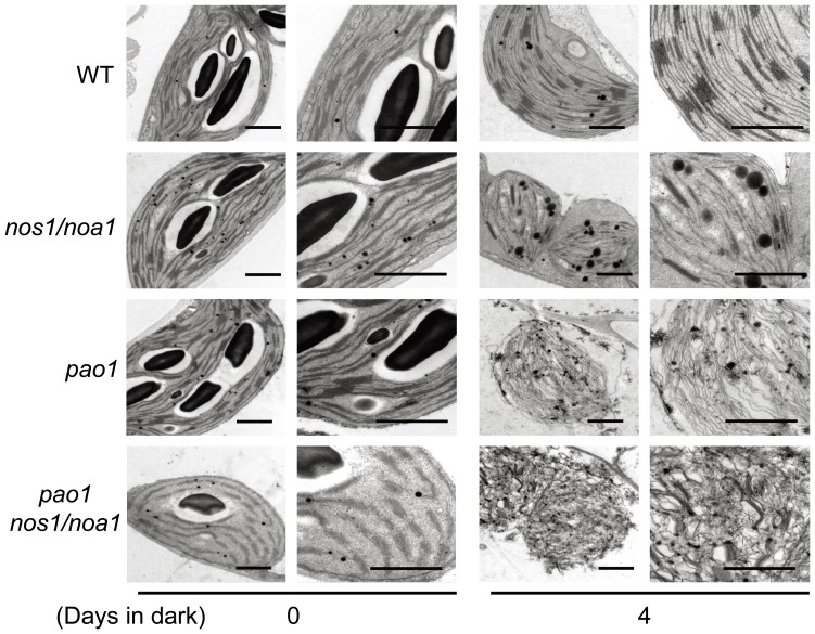
Figure 9. Chloroplast ultrastructures of detached leaves during dark-induced senescence.
Cross-sectional analysis of thylakoid membranes in chloroplasts from the fully-expanded leaves detached from wild type, nos1/noa1, pao1 and the double mutant pao1 nos1/noa1 plants after 0 d and 4 d of dark-treatment by transmission electron microscopy (TEM). Low magnification overview (left) and close-up images (right) of the chloroplast ultrastructure were shown respectively at day 0 and day 4. Bars = 1 µm.

