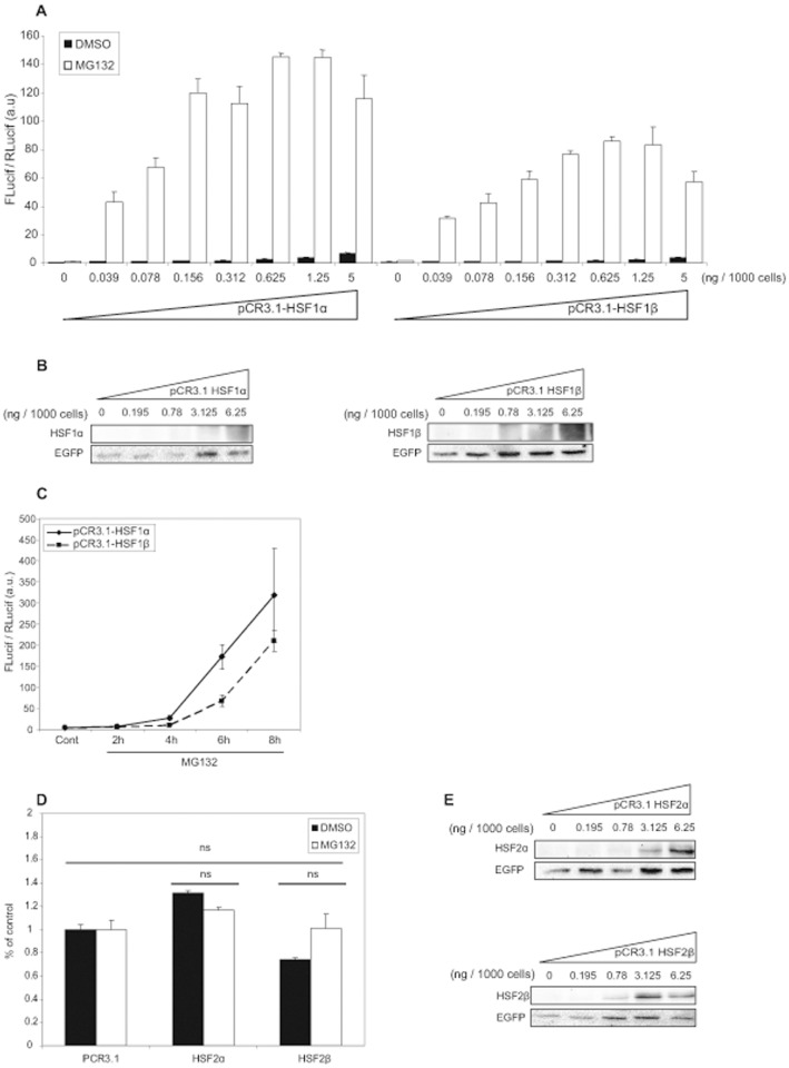
Figure 1. Transcriptional activity of HSF1 and HSF2 isoforms.
(A) Hsf1.2−/− iMEFs were co-transfected with increasing quantity of pCR3.1-HSF1α (left), or pCR3.1-HSF1β (right), in addition to pHSEx2-TATA-Luc used as a reporter gene. DNA quantities were adjusted with empty pCR3.1. Transfection efficiency was assessed using the pTK-Rluc reporter gene. Cells were treated with MG132 at 2.5 µM (white) or with DMSO (black) as control, for 8 h. Results correspond to the ratio between firefly luciferase (FLucif) and renilla luciferase (RLucif) activities. The data are from a representative experiment including three independent replicates (mean +/− SD). (B) Representative Western-blot showing the expression of HSF1 isoforms after transfection. Hsf1.2−/− iMEFs were co-transfected with increasing quantity of pCR3.1-HSF1α (left) or pCR3.1-HSF1β (right) and pEGFP as control for transfection efficiency. Cells were treated with MG132 at 2.5 µM and immunoblots for HSF1 and GFP were performed. (C) Hsf1.2−/− iMEFs were co-transfected with 12.5 ng of pCR3.1-HSF1α (full line), or pCR3.1-HSF1β (dotted line), with two reporter genes described previously. Cells were treated with MG132 at 2.5 µM or with DMSO, for 2 h, 4 h, 6 h or 8 h. Results correspond to the ratio between firefly luciferase (FLucif) and renilla luciferase (RLucif) activities and are the mean of three independent experiments +/− SD. (D) Hsf1.2−/− iMEFs were co-transfected with 12.5 ng of pCR3.1-HSF2α, or pCR3.1-HSF2β, with the reporter genes as described in (A). Cells were treated with MG132 at 2.5 µM (black), or with DMSO (white), for 8 h. Results are expressed in percentage of empty vector and represent the mean of three independent experiments +/− SD (Student’s t test, ns: no significant). (E) Representative Western-blot showing the expression of HSF2 isoforms after transfection. Hsf1.2−/− iMEFs were co-transfected with increasing quantity of pCR3.1-HSF2α (high panel) or pCR3.1-HSF2β (low panel) and pEGFP as control for efficiency. Cells were treated with MG132 at 2.5 µM and immunoblots for HSF2 and GFP were performed.

