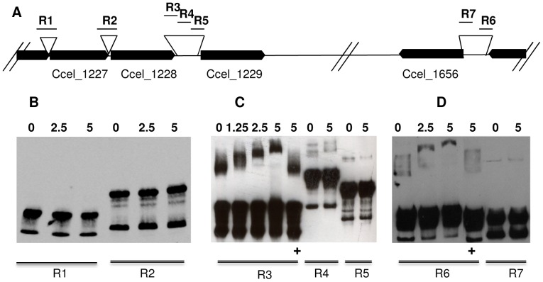
Figure 5. Binding of a truncated XydR regulator to target regions.
A) Schematic representation of targeted regions [R1 (309 bp), R2 (347 bp), R3 (272 bp), R4 (429 bp), R5 (345 bp), R6 (246 bp), and R7 (240 bp)] upstream genes at loci Ccel_1227, CCel_1228, Ccel_1229, and Ccel_1656. The loci of the different genes are represented as black arrows. B, C and D) Electromobility shift assays were carried out with 20 fmol of 3′ OH biotin-labeled regions (R1, R2, R3, R4, R5, R6, and R7) without any protein or with various amounts of purified MBP-Δ116 which are indicated in nM on top of the lanes. Competitive inhibition of the bindings were performed with 5 nM of MBP-Δ116, 20 fmol of 3′ OH biotin-labeled of regions R3 and R6 and 2 pmol of unlabeled R3 and R6 regions respectively (lanes with+at the bottom).

