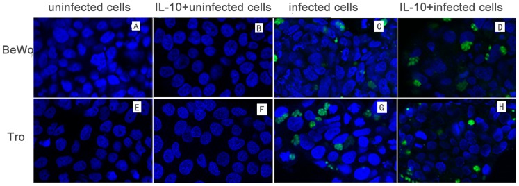
Figure 4. Hoechst 33258 staining of BeWo cells and trophoblasts at 24 hr after challenge with T. gondii.
(A) Uninfected BeWo cells, (E) uninfected trophoblasts, (B) uninfected BeWo cells treated with IL-10, (F) uninfected trophoblasts treated with IL-10, (C) infected BeWo cells, (G) infected trophoblasts, (D) infected BeWo cells treated with IL-10, and (H) infected trophoblasts treated with IL-10 were fixed, stained with Hoechst 33258, and observed by microscopy. Apoptosis was indicated by nuclei fragmentation or crescent shaped nuclei. Most of apoptotic nuclei were at a distance from the parasitophorous vacuoles (PV) and nuclei close to parasitophorous vacuoles usually showed normal appearances. The results also showed that the apoptotic cells increased in infected cells compared to uninfected cells and decreased with IL-10 treatment while there was no significantly difference between IL-10 treated uninfected cell and uninfected cells.

