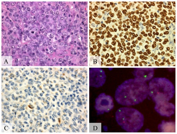
Figure 1. A20 monoallelic deletion in pyothorax-associated lymphoma.
(A) Diffuse proliferation of lymphoid cells, (hematoxylin-eosin stain, Olympus BX51, magnification ×200; inset ×400). (B) Positive signals in the nucleus of almost all tumor cells, (Epstein-Barr virus encoded RNA1, Olympus BX51, magnification ×400). (C) Positive signals in >50% of the tumor cells, (latent membrane protein-1, Olympus BX51, magnification ×400). (D) Monoallelic deletion of A20 detected by fluorescent in situ hybridization. A20 probe (orange) and chromosome 6 centromeric probe (green) (Olympus IX71, colors corrected after acquisition with Adobe Photoshop).

