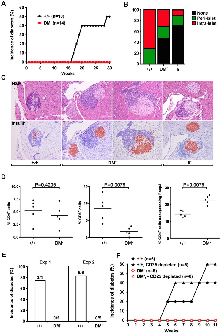
Figure 6. DM mutant NOD mice protected against autoimmune diabetes fail to develop pathogenic CD4+ T effectors.
(A) The onset of diabetes was evaluated by measurement of blood glucose levels in age-matched DM mutant and wildtype females. The incidence of diabetes has been absolutely zero over the past 2 years with parents in homozygous mutant breeding cages routinely tested at 6 mo of age. (B) The percentage of islets with a given degree of infiltration at 5 mo of age. (C) Typical examples of islet architecture assessed by H & E and insulin staining demonstrate substantial destruction by intra-islet infiltrates in wildtype mice, the normal islets predominantly seen in Ii chain mutants, and benign peri-islet infiltrates present in DM deficient NOD mice. (D) The representation of CD4, CD8, and Foxp3+ T cell subsets in pancreatic infiltrates was analyzed by flow cytometry. (E) DM-deficient NOD mice are resistant to cyclophosphamide-induced type 1 diabetes. Blood glucose levels were measured in age-matched wildtype and mutant females injected i.p. with cyclophosphamide at a dose of 200 mg/kg. (F) Depletion of CD25+ T cells fails to reveal the presence of pathogenic CD4+ T effectors in DM mutants. NOD.scid females were reconstituted with the indicated T cell populations.

