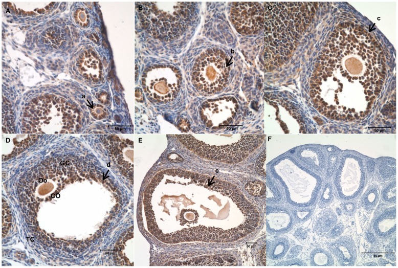
Figure 1. Expression of furin in follicles at different stages of folliculogenesis in the rat ovary.
Ovaries from PMSG (10 IU; 0, 24, or 48 h)-primed immature rats were processed for paraffin section. Furin was immunolocalized by immumohistochemistry. Sections from the forty-eight hour ovary were included as the negative control and no positive signals were detected. A–E: Follicles at different stages of development, A–B: the section of 0 h after PMSG injection, C–D: the section of 24 h after PMSG injection, E–F: the section of 48 h after PMSG injection, a: primary follicle, b: pre-antral follicle, c: early antral follicle, d: antral follicle, e: mature follicle; GC: granulosa cell, TC: theca cell, Oo: oocyte, CO: cumulus oophorus. F: negative control. Bar = 50 μm.

