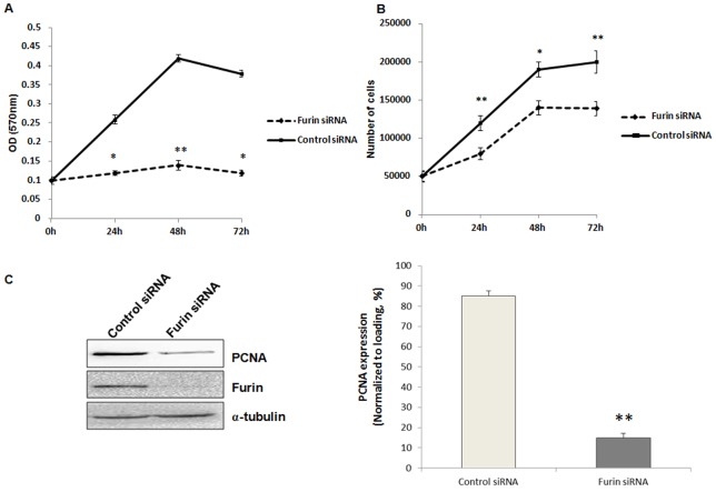
Figure 5. Proliferation of granulosa cells was inhibited by furin siRNA.
Granulosa cells were transfected with furin siRNA or contron siRNA for 48 h before being included in the proliferation assay. A. Cell proliferation was examined by MTT assay. Data represented means ± SD of OD (570 nm) at 0, 24, 48, 72 h of siRNA transfection (each concentration was tested in triplicate). *, P<0.05 and **, P<0.01 were significantly different from control values evaluated by t test. B. Cell numbers after 0, 24, 48, 72 h of siRNA transfection. C. Western blot analysis of PCNA expression in granulosa cells after transfection with furin siRNA for 48 h. α-tubulin was used as a loading control. Shown on the left were representative Western blot images and on the right was the statistical analysis of three independent experiments (**, P<0.01).

