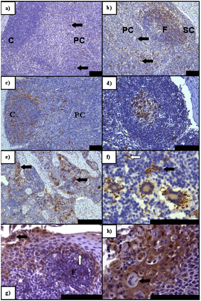
Figure 3. PPRV IHC on sections of lymphoid tissue taken at PME showing pertinent features of PPRV infection. a).
A greater degree of immunolabelling (arrows) is seen in the paracortex (PC) (arrows) of the RPLN than in the cortex (C) (5 dpi); b) Antigen distribution in the subcapsular layer and the follicles at 7 dpi in the MLN. Paracortical virus antigen still remains (arrows); c) Primarily cortical immunolabelling within the RPLN (9 dpi) with antigen also remaining within the PC; d) In contrast to b) the germinal center of this follicle within the MLN contains virus antigen that is absent from the follicular mantle (9 dpi); e) Intense immunolabelling within the LPSLN medulla (5 dpi) with extensive syncytia formation (arrows); f) Predominately peripheral paracortical immunolabelling of syncytia within the LPSLN (7 dpi). Dendritic-type cells also present and positive for virus antigen (arrow) with an infected lymphocyte also present (open arrow); g) Immunolabelling within pharyngeal tonsil (5 dpi) indicating early epithelial infection noted both basally (open arrow) adjacent to an infected lymphoid follicle (F) and apically, abutting the crypt lumen (solid arrow); h) Advanced epithelial infection of the pharyngeal tonsil (7 dpi) with syncytia formation. All scale bars represent 100 µm.

