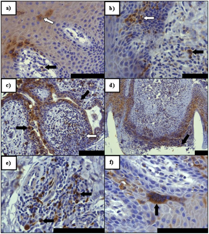
Figure 4. PPRV IHC on sections of facial epithelial tissue. a).
Nasal mucosal epithelium (7 dpi). Immunolabelling of lymphoid (arrow) and epithelial cells (open arrow) indicating early PPRV infection of the nasal lamina propria (LP) and mucosal epithelium. Epithelial immunolabelling is noted almost exclusively in basal layers, surrounding LP papillae, with extension into the stratum spinosum; b) Lingual mucosal epithelium (9 dpi) Immunolabelling indicative of early infection in both the basal epithelium and stratum spinosum (arrow) alongside positively labelled immune cells in the LP (open arrow); c) Conjunctival mucosal epithelium (9 dpi). Evidence of advanced epithelial and proprial infection involving a mixed population of inflammatory and epithelial cells around an exocrine gland (arrows). Note in particular the immunolabelling within the proprial lymphoid follicle circumscribed by this gland (open arrow); d) Nasal skin (9 dpi) - marked epithelial infection and erosion (arrow) in and around two hair follicles; e) Labial mucosal epithelium (9 dpi) Following epithelial infection, lymphoid follicles were often seen to have formed in the LP of facial mucosae. Here a large lumber of positively immunolabelled lymphoid cells are seen (arrows); f) Labial mucosal epithelium (9 dpi) with a large epithelial syncytium (arrow) seen in the lower stratum spinosum layer. All scale bars represent 100 µm.

