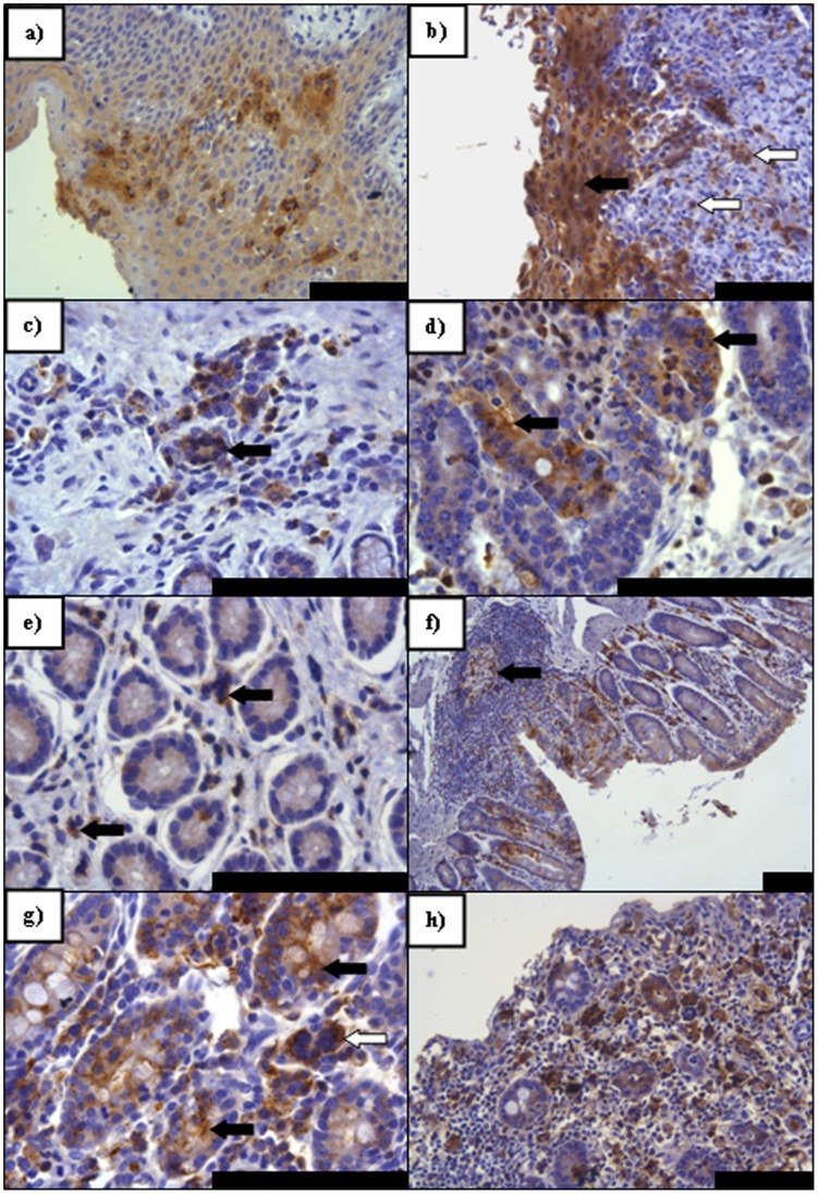
Figure 5. PPRV IHC on sections of digestive tract tissue taken at PME showing pertinent features of PPRV infection. a).
An isolated focus of virus antigen detected in the omasum (7 dpi) in an area of epithelial trauma; b) Marked immunolabelling both of the epithelial (arrow) and proprial cells (open arrows) within the oesophagus (9 dpi); c) Foci of infection within abomasal crypts amongst lymphoid cells including a small syncytium (arrow); d) Severe abomasal infection (9 dpi) of crypt epithelial cells (arrows); e) Positively labelled lymphocytes (arrows) disperse throughout the lamina propria (LP) of the rectum (9 dpi); f) A lymphoid aggregate in rectal epithelia (9 dpi) taking the form of a true follicle with a germinal centre (arrow) containing many positively immunolabelled lymphocytes; g) Positive immunolabelling in the caecum (7 dpi) seen abundantly in proprial lymphocytes and within caecal glands (arrows) alongside a lymphoid syncytium (open arrow); h) Marked viral infection of both glandular epithelial cells and the immune/inflammatory cells present within the caecum (9 dpi). All scale bars represent 100 µm.

