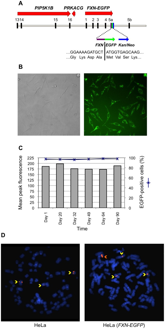
Figure 1. Characterization of HeLa (FXN-EGFP) stable cell lines.
(A) Diagrammatic representation of the BAC genomic DNA fragment containing the FXN-EGFP genomic reporter construct. The sequence includes exons 13–16 of the PIP5K1B gene and the PRKACG gene upstream of the FXN locus, and about 23 kb of additional sequence downstream of exon 5b. The exon 5a–EGFP–Kan/Neo region is shown in greater detail. (B) Microscopic imaging. Transmitted light (left) and fluorescence (right) images of a HeLa (FXN-EGFP) stable cell line. EGFP expression produced by the FXN-EGFP genomic reporter is evident in all cells. (C) Flow cytometric analysis. The levels of EGFP expression (left Y-axis) and the proportion of EGFP-positive cells (right Y-axis) were stable following growth in continuous culture. (D) Determination of transgenic fragment integration site by FISH. Rhodamine-labeled RP11-265B8 was hybridized onto metaphase chromosomes (DAPI stained) of HeLa (left) and HeLa (FXN-EGFP) (right) cells. Three hybridization signals (yellow arrows) corresponded to the endogenous FXN gene. The presence of one additional brighter signal (orange arrow) establishes the presence of a single integration site containing multiple copies of the FXN-EGFP transgene.

