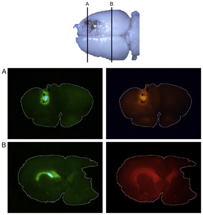
Fig. 2.
Ad5-green fluorescent protein (GFP) and rhodamine–dextran distribution in coronal rat brain slices. Ad5-GFP and rhodamine–dextran were co-infused intracranially in male rats. (A) GFP (green) expression is highest around the site of injection and rhodamine–dextran (red) is seen throughout the hemisphere. (B) At a more caudal slice, rhodamine–dextran shows greater spread across the corpus callosum. A smaller area of GFP expression is confined to the white matter.

