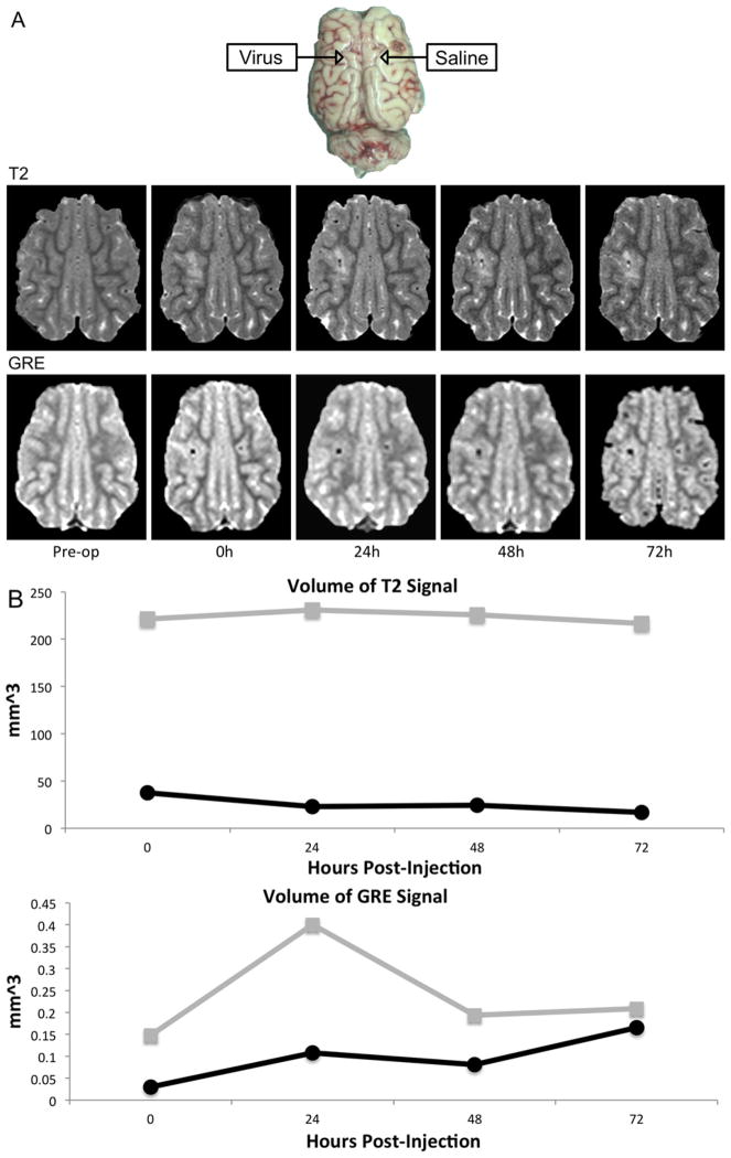
Fig. 6.
Post-injection imaging of super-paramagnetic iron oxide (Fe3O4) nanoparticle (SPION)-Ad5/3 cRGD and saline. Nanoparticle-labeled Ad5/3-cRGD-green fluorescent protein (GFP) was injected into subcortical white matter of a pig and contralateral saline injection was used as a control. (A) Axial (upper) T2-weighted MRI showing greater T2-weighted hyperintense area around the site of virus compared to saline injection that persists for up to 72 hours; and (lower) gradient echo (GRE) showing hypointensity at the virus site, and a smaller area of hypointensity on the saline-infused side. (B) The volume of T2 hyperintensity is greater with virus injection (gray) compared to saline injection (black) as measured by both T2-weighted and GRE signal.

