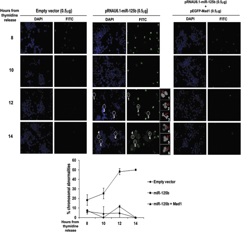
Figure 5.
miR-125b induces CIN. Chromosomal abnormalities are enhanced upon ectopic expression of miR-125b. UPCI:SCC084 cells were transiently transfected with either empty vector (0.5 μg), miR-125b expression plasmid (0.5 μg), or miR-125b expression plasmid (0.5 μg) and pEGFP-C1-Mad1 (0.5 μg) and synchronized. Immunostaining was done with antibody against p-H3 and nuclei were stained with DAPI at 8, 10, 12 and 14 h from second thymidine release. Cells were visualized under a fluorescence microscope. Representative images are shown and the defects are indicated by white circles in the full images and by red arrows in the cropped greyscale images (1–6). Lagging chromosomes can be visualized clearly in the cropped greyscale images 1, 2 and 3 (middle panel). Scale bar represents 500 μm. Images represent × 40 magnification. Percentages of mitotic abnormalities were calculated and plotted as shown. Data represents three independent experiments (N=3) and shown as average±S.D.

