Figure 6.
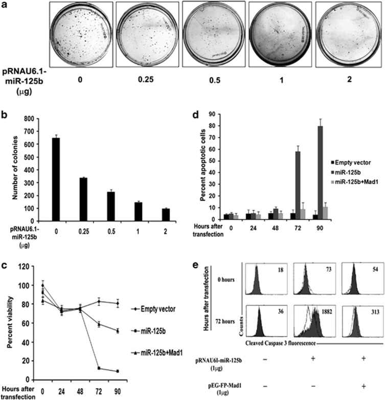
Excess miR-125b leads to cell death. (a) Clonogenicity of cells is affected by ectopic miR-125b. UPCI:SCC084 cells were seeded at a density of 103 and transiently transfected with 0, 0.25, 0.5, 1 and 2 μg miR-125b expression plasmid or empty vector. Colonies were stained with methylene blue after a week. (b) Colonies were counted from (a) and plotted as shown. (c) Ectopic miR-125b reduces cell viability. 105 UPCI:SCC084 cells were transiently transfected with either empty vector (1 μg), miR-125b expression plasmid (1 μg), or miR-125b expression plasmid (1 μg) and pEGFP-C1-Mad1 (1 μg) (N=3). MTT reagent was added at a final concentration of 50 μg/ml at 0, 24, 48, 72 and 90 h post-transfection Dye was dissolved in DMSO and read in a microplate reader. (d) Excess miR-125b induces apoptosis. 105 UPCI:SCC084 cells were transiently transfected with either empty vector (1 μg), miR-125b expression plasmid (1 μg), or miR-125b expression plasmid (1 μg) and pEGFP-C1-Mad1 (1 μg). At 0, 24, 48, 72, 90 h post-transfection, cells were subjected to annexin V-FITC/PI staining followed by flow cytometry analysis. (e) Ectopic miR-125b increases cleaved caspase-3 levels. 105 UPCI:SCC084 cells were transiently transfected with either empty vector (1 μg), miR-125b expression plasmid (1 μg), or miR-125b expression plasmid (1 μg) and pEGFP-C1-Mad1 (1 μg). Cells were fixed, permeabilized and stained with control antibodies or cleaved caspase-3 antibody at 0 and 72 h post-transfection followed by flow cytometric analysis. Open and filled histograms represent staining with normal rabbit serum and anti-cleaved caspase-3 antibody, respectively. Values within histograms represent specific mean fluorescence intensity upon subtracting respective control values. For (b–d), data represent three independent experiments (N=3) and shown as average±S.D. For (a) and (e), images are representative of three independent experiments

