
Figure 2.
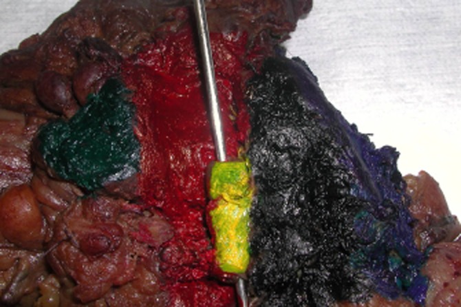
Pancreaticoduodenectomy (standard) specimen (the same case as previous Fig. 1, view from the closer distance). Posterior view (mapping using different colours/dyes). Green, distal pancreatic resection margin; Red, superior mesenteric vein (vascular groove) dissection margin; Yellow, superior mesenteric vein (SMV) adhered to the posterior lateral aspect of the SMV dissection margin (the probe inserted in the lumen of the SMV); Black, SMA resection margin; Blue, posterior surface of the uncinate process dissection margin