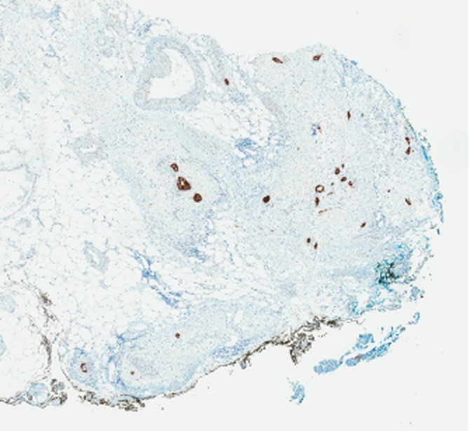
Figure 3.

Invasion of the peripancreatic adipose tissue by single malignant glands (black coloured structures highlighted by keratin stain AE1/AE3) along the SMA margin, marked by black ink, sectioned perpendicular to the resection margin (see standardized protocol). AE1/AE3 keratin stain, original magnification 2×
