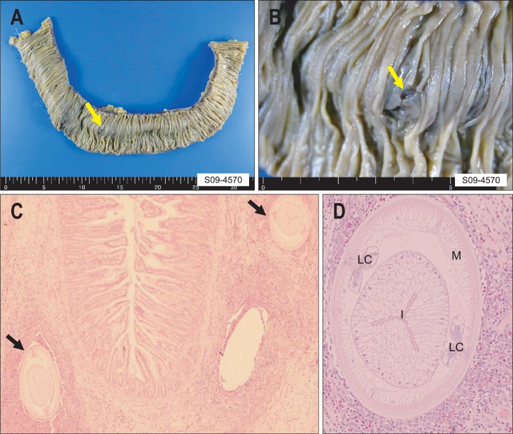
Fig. 3.
Small bowel segmental resection in a patient with small bowel anisakiasis. (A, B) The resected small bowel specimen (approximately 30 cm) showing the penetration of an Anisakis larva into the bowel wall (yellow arrow). (C) H&E-stained cross section showing two larvae (black arrows) surrounded by a thick cuff of acute inflammatory cells with numerous eosinophils (×200). (D) High-power view of a cross section through a larva (H&E stain, ×400).
M, muscle layer; LC, lateral chord; I, intestine.

