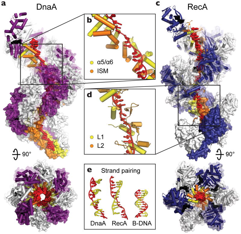
Figure 3.
DnaA binds single-stranded DNA in a manner similar to RecA. (a) Side and top views of oligomerized DnaA (PDB ID: 3R8F), showing twelve DnaA subunits, each colored white or purple, complexed with DNA (red). AMPPCP and Mg2+ are shown as black spheres and the DNA engagement elements, helices α5/α6 and the ISM, are colored yellow and orange, respectively. (b) Close-up of the DnaA oligomer bound to the backbone of DNA. (c) Side and top views of a RecA filament, constructed by aligning four copies of a RecA pentamer model (PDB ID: 3CMW [69]). Twelve RecA subunits, each colored white or blue, are shown complexed with DNA (red). ADP•AlF4 and Mg2+ are shown as black spheres and the DNA engagement elements, loops L1 and L2, are colored yellow and orange, respectively. (d) Close-up of the RecA oligomer bound to the backbone of DNA. Both DnaA and RecA bind DNA in an extended conformation with large gaps between trinucleotide base stacks. (e) (left) Cartoon model showing how complementary base triplets (yellow) would pair (in a B-DNA manner) with single-stranded-DNA bound to DnaA (red). Formation of a continuous base-paired strand is prevented by the orientation of successive DnaA-bound triplets. (middle) Same DNA view, but as seen in RecA, which orients triplets to allow pairing of an extended complementary strand for promoting duplex formation and strand exchange (PDB ID: 3CMX [69]). (right) B-DNA.

