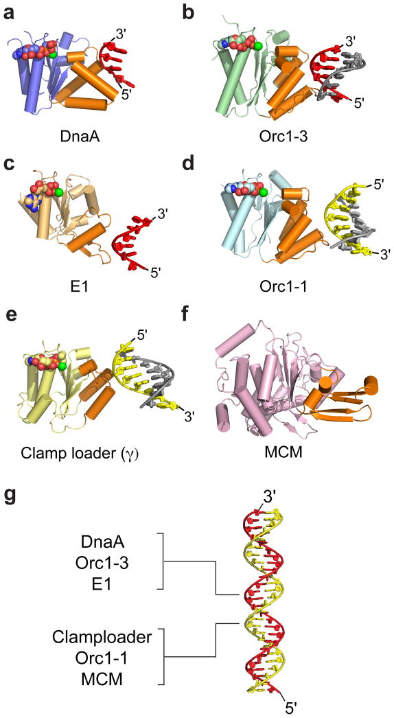
Figure 4.
DNA recognition by replicative AAA+ proteins. Nucleic acid substrates are all engaged using structural elements that reside on the same face of the AAA+ domain (orange). (a) Bacterial initiator DnaA (AAA+ domain, blue) bound to single-stranded DNA [9]. (b) Archaeal initiator Orc1-3 (AAA+ domain, green) bound to duplex DNA (PDB ID: 2QBY [62]). (c) Viral initiator E1 (AAA+ domain, light orange) bound to single-stranded DNA (PDB ID: 2GXA [71]). (d) Archaeal initiator Orc1-1 (AAA+ domain, cyan) bound to duplex DNA (PDB ID: 2QBY). (e) Bacterial clamp-loader subunit γ (AAA+ domain, yellow) bound to primer-template DNA (PDB ID: 3GLF [72]). (f) Archaeal Mcm (AAA+ domain, pink) (PDB ID: 3F9V [80]); although a DNA-complex has yet to emerge for this enzyme, mutagenesis studies have implicated the highlighted regions in binding substrate and helicase activity [81,82]. (g) Duplex DNA (red/yellow) with the strand bound by various AAA+ proteins indicated. For panels a–f, DNA is shown as red/grey or yellow/gray cartoons. Nucleotide co-factors are represented as spheres colored by atom.

