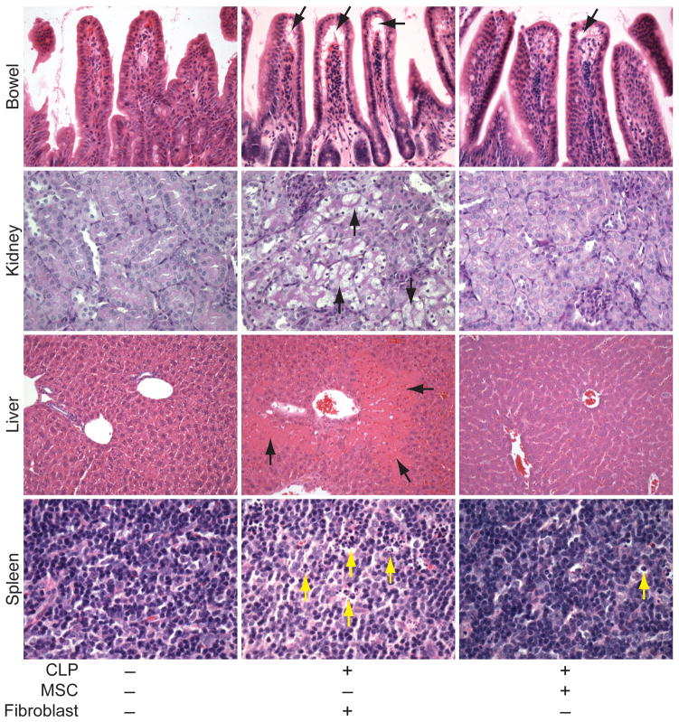
Figure 5.
Representative images of organs from mice that received MSCs or fibroblasts after the onset of CLP-induced sepsis. H&E staining of sections from small bowel, liver, and spleen, and PAS staining of kidney, from mice 24 hours after sham (−) or CLP (+) surgery. In the CLP mice, 2 hours after the onset of sepsis, MSCs or fibroblasts were administered. The arrows point to subepithelial spaces in the villi (small bowel, black arrows), tubular epithelial cell swelling (kidney, black arrows), necrotic regions (liver, black arrows), and tingible body macrophages, predominantly in the white pulp, which have phagocytized apoptotic cells (spleen, yellow arrows). Light microscopy images, small bowel and kidney (20X objective), liver (10X objective), and spleen (40X objective).

