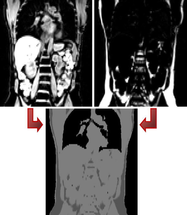
Fig. 5.

Dixon-based segmentation for whole-body attenuation correction shown in [44] (courtesy of A. Martinez-Moeller): MRI water (top left) and fat (top right) images acquired with a 2-point Dixon sequence are combined and segmented to generate the attenuation map for lungs, adipose tissue, soft tissue, and background
