Figure 1.
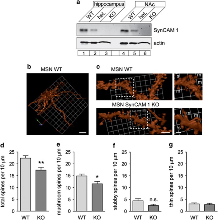
SynCAM 1 is expressed in NAc and the absence of SynCAM 1 decreases synapse density. (a) SynCAM 1 expression was tested by immunoblotting of equal protein amounts of the indicated tissues from WT, heterozygotic (het.), and SynCAM 1 KO mice, which served as a negative control for the antibody. Actin was used as a loading control. (b) Three-dimensional reconstruction of a MSN in WT NAc. The neuron was visualized after biolistic transfer of the dye DiI. No gross morphological differences were observed between MSN from WT and SynCAM 1 KO mice. Scale bar, 20 μm. (c) Left, dendritic MSN segments in NAc from WT (top) and SynCAM 1 KO (bottom) male mice. Dendrites were visualized as in (b). Scale bar, 5 μm. Right, enlarged view of boxed areas. Labels mark m, mushroom; s, stubby; t, thin spines. Scale bar, 2 μm. (d–g) Lower density of total spines (d) and mushroom-type spines (e) in MSN of NAc from SynCAM 1 KO. The trend towards lower stubby spine density in the KO was not significant (f). Thin spines were unaffected (g). Data were obtained from male littermate mice imaged as in (c), with the experimenter blind to condition. (n=23 neurons from six WT mice, 10 neurons from three KO mice) *P<0.05; n.s., not significant.

