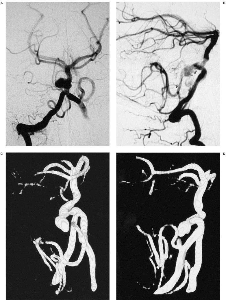
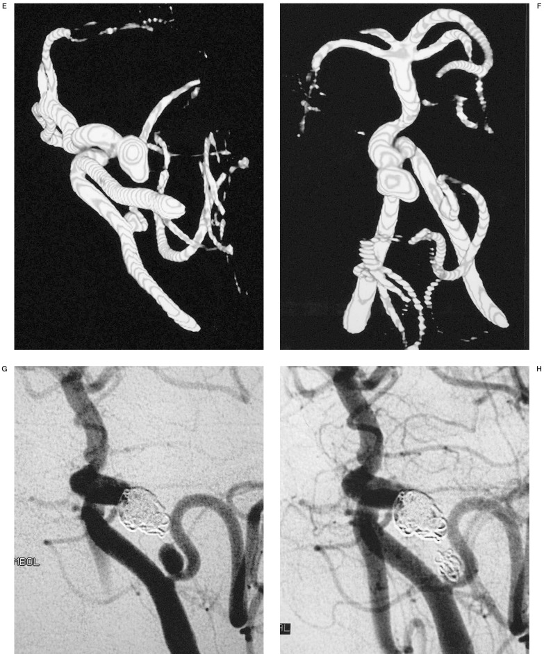
Figure 5.
The three-dimensional nature of this technique and the ability to view the three-dimensional imaging volume at multiple angles was helpful in identifying the relationship of this aneurysm of the basilar artery and evaluates the neck of the aneurysm beneath the overlapping tortuous basilar artery and demonstrates a second aneurysm of the posterior inferior cerebellar artery. This ability to depict the aneurysmal neck and the projection of the aneurysm relative to the parent artery is advantageous for evaluating the outcome of clipping or embolization of the aneurysm. In this particular case, this information advocates the best protection and the correct choice of initial coil to build a basket permitting total occlusion of the aneurysm.


