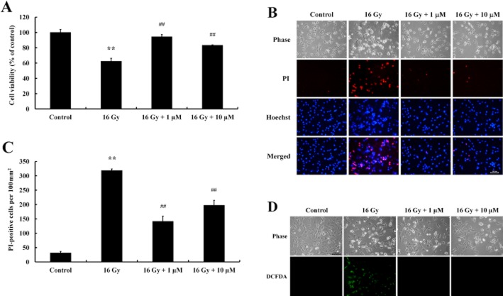
Figure 1.
Protective effect of baicalein on irradiation-induced cell death in C17.2 NPCs. Cells were seeded and cultured for 24 h, treated with baicalein (1 μM, 10 μM), and exposed to 16 Gy 12 h later. (A) After incubation for 24 h, cell viabilities were determined by MTT assay. Analysis showed exposure to radiation significantly decreased cell viability and that baicalein had a protective effect. Data shown are means ± SE (n = 4 cultures per group). **P < 0.01 versus controls; ##P < 0.01 versus 16 Gy (anova using Fisher's PLSD procedure). (B) Irradiation-induced cell death was assessed by nuclear staining with Hoechst 33342 and PI. Increased numbers of PI-stained cells were observed after irradiation. Scale bar = 100 μm. (C) PI-stained cells were quantified by counting under a fluorescence microscope. Data shown are means ± SE (n = 4 cultures per group). **P < 0.01 versus Controls; ##P < 0.01 versus 16 Gy (anova using Fisher's PLSD procedure). (D) Irradiation-induced ROS generation was detected by DCFDA fluorescence. Cells were treated with baicalein (1 μM, 10 μM) for 12 h, exposed at 16 Gy for 24 h, and intracellular ROS was detected by DCFDA staining under a fluorescence microscope. Scale bar = 100 μm.

