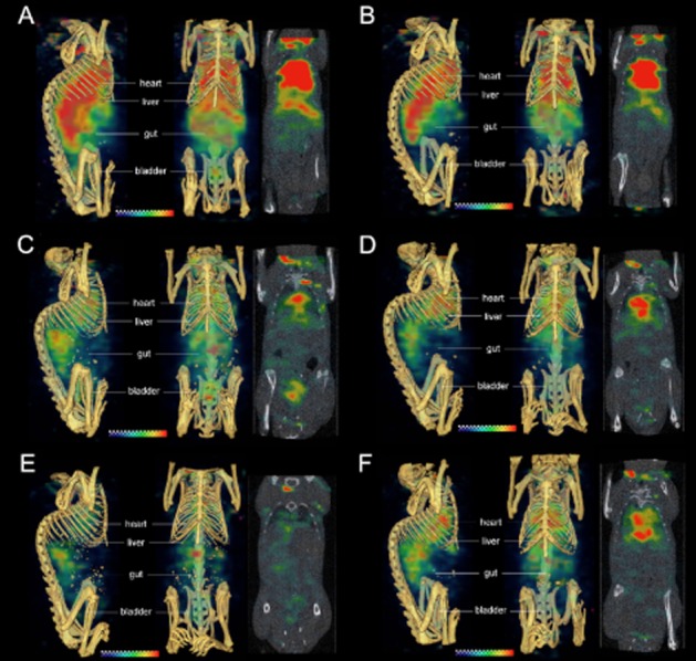
Figure 6.

SPECT-CT fusion images of non-tumour-bearing SCID mice receiving [111In]-anti-TENB2-MMAE (ADC) at tracer only (A, C, E) or 10 mg·kg−1 (B, D, F) dose levels at 3, 48 and 72 h after i.v. injection (A–B, C–D and E–F respectively). Three-dimensional volume rendering images from sagittal (left) and coronal (centre) perspectives and coronal tomographic images (right) are shown in each panel. Note that the high signals above the thorax and below the animals' heads are both caused by reconstruction edge artefacts that are more severe at early time points when higher levels of radioactivity remain in the blood pool.
