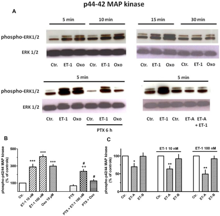
Figure 15.
Western blot analysis of phospho-p42/44 (ERK1/2) MAPK in MRC-5 human lung fibroblasts. Cells were cultured in 55 mm dishes to nearly confluence, serum-starved for 24 h and exposed to 100 nM ET-1, 10 μM oxotremorine (Oxo), or vehicle (Ctr.) for 5–30 min (as indicated, A) or 5 min (B and C). In the respective experiments, pertussis toxin (PTX, 50 ng·mg−1) was present 6 h and the ET-A (BQ123, 1 μM) or ET-B (BQ788, 100 nM) receptor antagonist 30 min prior to exposure to ET-1. In controls, cells were treated with solvent at the respective time points (i.e. 5 min and additionally 30 min or 6 h before cell lysis respectively). Cell lysates were prepared, and 25 μg was separated in 4–10% acrylamide Tris–glycine gels, immobilized to PVDF membranes and detected by antibodies specific for phosphorylated and unphosphorylated ERK. Densitometrical quantification of the p44/42 bands was performed, and values (arbitrary units, normalized over unphosphorylated ERK) are expressed as % of the respective control, in absence or presence of ET-1 and absence or presence of the solvent DMSO (which was required to dissolve BQ788). Given are means ± SEM of n = 4–14. Significance of differences: *P < 0.05, **P < 0.01, ***P < 0.001 versus respective Ctr.; #P < 0.01 versus respective value in absence of PTX; ++P < 0.05 versus respective value in presence of PTX alone.

