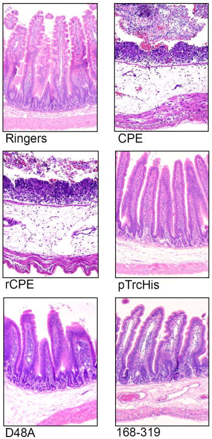
Figure 3.

Histopathological changes induced by the toxin preparations described in Figure 2, in rabbit small intestinal loops. After the 6 h fluid accumulation experiments, rabbit small intestinal loops selected for histological examination were formalin-fixed and embedded in paraffin. Hematoxylin and eosin was used to stain 4 μm thick sections of intestinal tissue treated with the indicated construct. The preparation used to treat each loop is noted at the bottom of each panel. Final magnification: 200×. Copyright © American Society for Microbiology, Infection and Immunity, 76, 2008, 3793-3800, DOI: 10.1128/IAI.00460-08. Used with permission.
