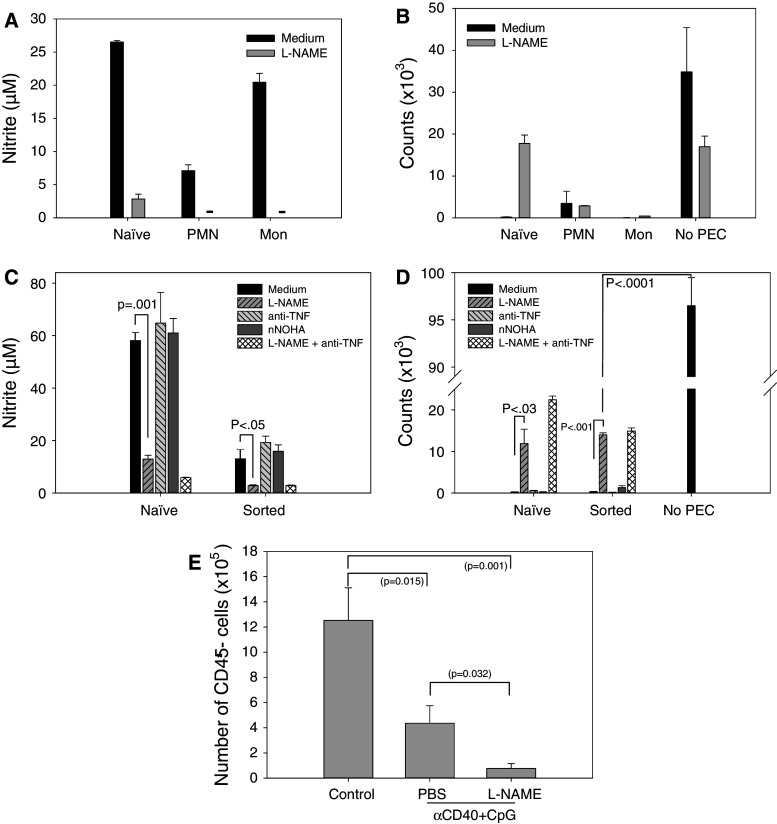
Fig. 6.
Role of NO, arginase and TNFα in the antitumor effects mediated by myeloid cells. PEC were obtained 15 (a, b) or 14 (c, d) days following injecting C57BL/6 mice i.p. with 105 B16 cells. PEC were sorted based on their CD11b+Gr-1+ phenotype (a–d) and also forward-side scatter characteristics, resulting in populations enriched for polymorphonuclear cells (PMN) and monocytes (Mon) (a, b) as also was verified by histology (not shown). PEC from naïve mice were used as the control. The inhibitors of NO, arginase and TNFα were added as described in “Materials and methods.” The functional data show mean ± SEM of nitrite levels produced by IFN-γ + LPS-stimulated sorted cell fractions (a, c) and the ability of the sorted cells to inhibit proliferation of B16 cells in vitro (b, d). Dashes signify values below detection limit (a). e Tumor-bearing mice were treated with anti-CD40 on day 11 and CpG on day 14 post tumor cell implantation. To neutralize NO in vivo, L-NAME was injected either i.p. at the dose of 50 mg/kg twice a day or given in the drinking water at the dose of 0.5 g/l on days 11–14. PEC were isolated on day 15, and the percentage of B16 tumor cells (CD45− PEC) was determined by flow cytometry. The combined data of two experiments are presented. Mean ± SEM of 10 mice per group or 7 mice for the last group (3 of 5 mice treated with anti-CD40/CpG/L-NAME died from toxicity following i.p. injections)

