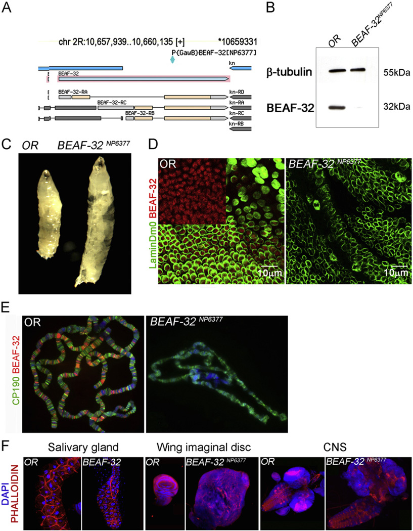
Fig. 1.
Mutations in BEAF-32 result in neoplastic growth. (A) Diagram showing the BEAF-32 locus adapted from the Flybase genome browser. The location of the P{GAW} transposon insertion is indicated; this insertion disrupts an exon common to both BEAF-32A and BEAF-32B isoforms. (B) Western blot for BEAF-32 (−32 kDa) showing the levels of this protein in wild type and mutant imaginal disc tissue; tubulin (55 kDa) was used as loading control. (C) Wild type and BEAF-32NP6377 mutant larvae showing the dramatic overgrowth of the later at the third instar stage. (D) Immunofluorescence microscopy using antibodies to the BEAF-32 protein in imaginal tissue; BEAF-32 (red) is present in nuclear foci while the peripheral location of lamin Dm0 (green) marks the nuclear lamina. (E) Immunofluorescence microscopy of polytene chromosomes showing the location of CP190 (green) and BEAF-32 (red). (F) Effect of BEAF-32 mutations in developing tissues such as salivary glands, wing imaginal discs and central nervous system. (For interpretation of the references to color in this figure legend, the reader is referred to the web version of this article.)

