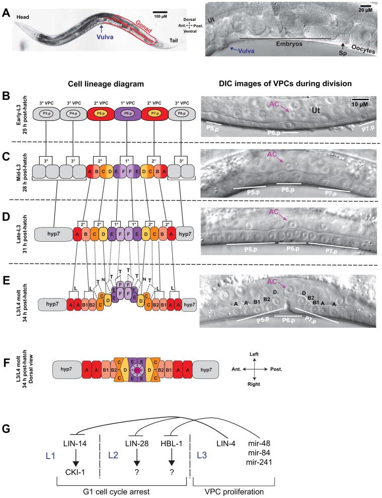
Figure 1.
Vulval cell divisions. (A) Adult worm at low (left) and high (right) magnification showing components of the reproductive system. Oocytes in the gonad (outlined in red) move in a distal to proximal direction and are fertilized in the spermatheca (Sp). Embryos pass into the uterus (Ut) and undergo the first rounds of cell division. Muscle contractions push embryos through the vulva to the external environment, where embryogenesis continues until hatching. The posterior gonad is shown; the anterior gonad has an identical arrangement. For additional images of the C. elegans reproductive system, see WormAtlas 14. (B–E) Cell divisions in the L3 larval stage. Left is schematic diagram; right is differential interference microscopy (DIC) images of live animals. Lateral view with anterior left and dorsal top. (B) The P6.p cell is induced to the 1° VPC fate, P5.p and P7.p are induced to the 2° VPC fate, and P3.p, P4.p, and P8.p take on the non-vulval 3° VPC fate. The anchor cell (AC) is located in the uterine epithelium (Ut) and positioned dorsal to P6.p (right DIC image). (C) All six VPCs divide in the mid-L3 larval stage, after which the 3° VPCs arrest cell divisions and initiate fusion with the hyp7 syncytium. The 1° VPCs differentiate to vulE and vulF cell types; the 2° VPCs differentiate to vulA, vulB, vulC, and vulD cell types. (D) The third VPC divisions take place in mid to late L3 and produces 12 cells in a longitudinal array. (E) The final VPC divisions occur during the L3/L4 molt. vulC, vulE, and vulF cell divisions occur in the transverse (T) orientation, which is indicated by dashed lines in the lineage diagram. vulA and vulB divide in the longitudinal orientation (L), and vulD does not divide (N). In the DIC image, only the longitudinally divided cells and vulD are in the plane of focus. (F) Dorsal view of the vulval primordium following terminal cell divisions, with the AC at the vulval apex. Modeled after 8. For additional schematics of vulval development and a time series of vulval images from the L3/L4 molt to adulthood, see WormAtlas 14. (G) Heterchronic genes regulate the timing of VPC divisions. LIN-14, acting through the cell cycle repressor cki-1, inhibits cell divisions in L1, and LIN-28 and HBL-1 inhibit L2 cell divisions, acting through unknown targets. In L3, the miRNA lin-4 represses lin-14 and lin-28 mRNA translation to promote cell division. Analogously, the miRNAs mir-48, mir-84, and mir-241 redundantly repress hbl-1 translation.

