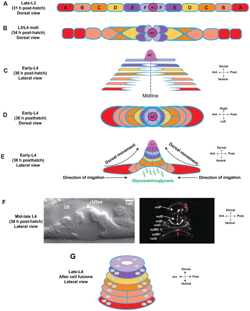
Figure 2.
Vulval invagination and toroid formation. (A) After the second cell divisions, the VPCs are arranged as a longitudinal array. The anchor cell (AC) is positioned at the junction of the vulF cells at the vulval midline. Apical domains are in blue here and in other panels. (B) Concurrent with the final cell divisions, the 1° VPCs invaginate by moving dorsally. vulF occupies the most dorsal position, with vulE positioned ventral to it. (C) VPCs extend apical membrane processes toward the midline and establish junctions with homotypic cells from the contralateral half, forming rings that encircle a tube in the center of the vulval primordium. In this schematic, vulC–vulF have formed rings. Sister vulA cells on each lateral half fuse shortly after the final cell division 8, shown as a single cell boundary. (D) Dorsal view of early-L4 VPCs, showing the lateral migration of vulval cells toward the midline. (E) Invagination is driven by distal VPCs migrating toward the midline along the ventral surface of inner cells, which forces cells to move dorsally 9. Glycosaminoglycans are expressed by the VPCs and are thought to maintain and shape the forming lumen 10, 11. (F) As the L4 stage continues, vulval cells continue to migrate and homotypic cells undergo fusions. A uterine lumen forms, and the uterine and vulval epithelia become separated by the thin membrane of the utse syncytium (left DIC image). The right image shows labeling of apical junctions with the apical membrane marker AJM-1::GFP 134. At this stage, the sister cells on each lateral half have fused, and contralateral vulA and vulD cells have fused at the midline. Red arrows indicate apical borders between vulB1, vulB2, vulC, vulE, and vulF cells. (G) Schematic of vulval toroids in late-L4 after fusion has completed. Grey circles represent nuclei. All homotypic cells use except vulB1 and vulB2. For additional images, see refs. 8, 9 and WormAtlas 14.

